
Patient Presentation:
An otherwise healthy 76-year-old woman was found on screening mammogram to have a new left breast mass. After her workup confirmed a small left breast tumor with no evidence of lymph node involvement, she had a lumpectomy and sentinel lymph node biopsy. Pathology showed a 1.1 cm infiltrating ductal carcinoma, grade I, ER/PR >90% and HER2 negative. Surgical margins were 3mm or greater, and there was no lymphovascular invasion. Two sentinel nodes were negative.
The patient recovered well from surgery and preferred to pursue both endocrine therapy and radiotherapy. For her radiotherapy, she was also interested in choosing a short, convenient approach that would allow her to maintain her busy schedule. Based on her clinical presentation, she met suitability criteria for partial breast irradiation by ASTRO consensus guidelines. She has C cup breasts, and the lumpectomy cavity was centrally located. Based on this anatomy, we considered both photon and proton approaches to partial breast radiotherapy, with the goal of minimizing radiation exposure to the heart, lungs and uninvolved breast tissue. While she is in excellent health, she has a strong family history of cardiac disease, which is an established risk factor for radiation-induced cardiac morbidity, depending on the dose of radiotherapy to the heart. She received proton therapy to maximally avoid the heart, which was a high priority for her.
Treatment:
She was treated with partial breast radiotherapy using protons on our proton registry study. Patients are candidates for this study when ASTRO suitability criteria for partial breast irradiation are met and patients agree to be treated on the registry study. We use a three-treatment partial breast irradiation approach, which was initially developed at the Mayo Clinic. The dose is 21.9 Gy in three treatments of 7.3 Gy each. Treatments are delivered over three consecutive days. We follow strict treatment planning criteria, which avoid radiation exposure to the uninvolved breast tissue (no more than 30% of the total breast volume can receive the prescribed dose), the heart (average heart dose must be
Outcome:
The patient tolerated radiotherapy well. She was able to tolerate the treatment position without difficulty and reported no acute toxicities. On follow-up two weeks after treatment, she had developed very mild erythema in the treated area of the breast but had no skin peeling and no breast pain or swelling. The erythema resolved in about two more weeks. She will require longer term follow-up to determine the ultimate cosmetic result and to assess for later radiation toxicities such as tissue fibrosis.
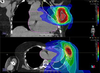
Figure 1 depicts coronal and axial images of her treatment plan. The radiation dose distribution shows complete avoidance of the heart and pericardium with protons (LEFT) versus photons (RIGHT). Notably, even with photons, radiation exposure to the heart is quite low. The improvement in radiation exposure to the heart with protons versus photons will vary significantly with the location of the target area in the breast and the patient’s unique anatomy.

Learn more about the Johns Hopkins Kimmel Cancer Center in the Washington D.C. Region at hopkinscancerdc.org.