Cardiovascular Report
November 1, 2013

Finch’s wife called 911, and he was rushed to a local community hospital. From there, he was airlifted to The Johns Hopkins Hospital. “When I arrived, they were ready. There was a whole team there to treat me and everything moved very fast,” says Finch.
The Johns Hopkins stroke team—including a stroke neurologist, an interventional neuroradiologist, technologists and nurses—is activated to meet an arriving stroke patient in the Emergency Department. Johns Hopkins is one of only about four dozen hospitals in the United States recognized by the Joint Commission as Comprehensive Stroke Centers.
“The designation means that a hospital has specialized staff and state-of-the-art technology available 24/7 to rapidly evaluate patients and provide them with the most advanced treatments and highest standard of care,” says Gordon Tomaselli, director of cardiology and past president of the American Heart Association, which launched the Comprehensive Stroke Center program with the Joint Commission in 2012.
Since Finch went to sleep at 11 p.m. without stroke symptoms that July evening, by the time he arrived at Johns Hopkins, he was outside the window of treatment with IV r-tPA. He was also pushing the eight-hour limit for intra-arterial therapy to aspirate the clot. Beyond eight hours, there is a risk that opening the vessel might actually cause more harm than good with a possible risk of converting an ischemic stroke into a hemorrhagic one, or there may be significant edema.
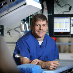
“We do treat patients beyond eight hours when we can get an MRI and the findings suggest that there is brain that can be salvaged,” says interventional neuroradiologist Martin Radvany. “In this case, the patient had a pacemaker, and we could not get an MRI, so we had to make decisions based on the CT scan and clinical judgment.”
When Radvany performed an angiogram, he saw that Finch’s distal right internal carotid artery was occluded.
“Because of his age and the fact that he was hemiplegic on his left side, we decided to move ahead with a mechanical thrombectomy,” says Radvany.
“We advanced an aspiration catheter into his artery, removed the clot and opened the vessel,” says Radvany. “We always have an anesthesia team present for these procedures, but since Mr. Finch was able to stay still for the procedure, we did it while he was awake with minimal sedation. This enabled us to assess his neurological status during the procedure.”
Finch says that 45 minutes after the procedure, he was able to move his left arm. After discharge from the hospital, he went to a rehabilitation facility, and six days later, he was once again walking on his own.
“It’s pretty amazing,” says Finch. “I’m still having a bit of difficulty with my handwriting but it’s getting better and I’m regaining my endurance and stamina. I don’t think I’d be where I am now in my recovery if things had happened differently.”

